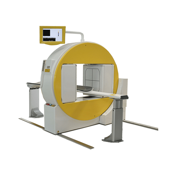
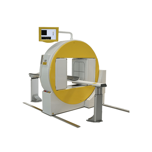
Gamma Camera and SPECT
Gamma cameras are based on the detection of gamma rays emitted from radionuclides. Radionuclides can be ingested or injected into the body. The camera accumulates counts of gamma photons, which are detected by crystals in the camera. Just like an X-ray, the gamma camera will yield a two-dimensional projection of a three-dimensional object. A tomographic version of the gamma camera is called SPECT, which yields slices through the body. Because the detection techniques of gamma cameras and SPECT are based on the same concept, the same radioisotopes can be used for both techniques. Commonly used isotopes include technetium-99m, iodine-123, and indium-111.
Scintigraphy (“scint”) is the use of gamma cameras to capture emitted radiation from internal radioisotopes to create two-dimensional images.
SPECT (single photon emission computed tomography) imaging, as used in nuclear cardiac stress testing, is performed using gamma cameras. Usually one, two or three detectors or heads, are slowly rotated around the patient’s torso.
Multi-headed gamma cameras can also be used for positron emission tomography (PET) scanning, provided that their hardware and software can be configured to detect “coincidences” (near simultaneous events on 2 different heads). Gamma camera PET is markedly inferior to PET imaging with a purpose designed PET scanner, as the scintillator crystal has poor sensitivity for the high-energy annihilation photons, and the detector area is significantly smaller. However, given the low cost of a gamma camera and its additional flexibility compared to a dedicated PET scanner, this technique is useful where the expense and resource implications of a PET scanner cannot be justified